Technical Guide: Building the MCAO Ischemia Model in Rats

The middle cerebral artery occlusion (MCAO) model is one of the most widely used and clinically relevant experimental paradigms in translational stroke research. By transiently occluding the middle cerebral artery with an intraluminal suture, researchers can reproduce focal cerebral ischemia with a well-defined infarct territory, allowing reliable evaluation of neuroprotective agents, thrombolytic strategies and post-stroke rehabilitation protocols. Reproducibility of the lesion depends heavily on consistent surgical technique, standardized occlusion time and meticulous peri-operative care, which this guide addresses step by step.
Animal Preparation
Healthy adult male Sprague-Dawley rats weighing 260–300 g are the standard choice for the intraluminal MCAO model. Before surgery, house animals under a 12 h light/dark cycle with free access to food and water, and allow at least 7 days of acclimatization after transport. On the day of surgery, fast the animals for 6–8 h (water ad libitum) to reduce gastric distension that may compromise breathing during anesthesia.
- Record baseline body weight and pre-operative neurologic status for each animal.
- Induce anesthesia with 3–4% isoflurane in 70/30 N2O/O2 and maintain at 1.5–2% during the procedure.
- Place the rat in a supine position on a heated pad (rectal temperature maintained at 37.0 ± 0.5 °C).
- Sterilize the ventral neck area with 70% ethanol and apply ophthalmic ointment to protect the eyes.
Surgical Procedure
Perform the surgery under a stereo-microscope with the rat positioned supine. Use blunt dissection to avoid trauma to the vagus nerve and carotid sheath structures.
- Make a midline neck incision and bluntly separate the strap muscles to expose the right common carotid artery (CCA).
- Carefully dissect the right external carotid artery (ECA) and internal carotid artery (ICA), ligating the ECA and its branches with 6-0 silk suture.
- Introduce a 4-0 or 3-0 monofilament nylon suture with a silicon-coated tip (18–22 mm) into the ICA lumen via the ECA stump.
- Advance the filament approximately 18–20 mm from the bifurcation until mild resistance is felt, confirming occlusion of the MCA origin.
- Secure the filament in place and occlude for the target duration (typically 60–120 min for transient ischemia).
- Withdraw the filament gently to allow reperfusion, then ligate the ECA stump and close the incision.
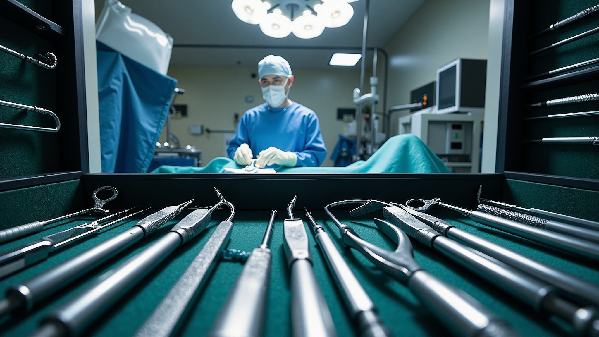
The intraluminal filament is the critical determinant of infarct size and consistency; verify that the silicone-coated tip is uniform in diameter and free of burrs before each use, and record the occlusion distance for every animal.
Post-operative Care
Standardized recovery management directly influences mortality and infarct variability. Transfer animals to a warm recovery cage immediately after surgery and monitor them closely during the first 24 h.
- Maintain body temperature at 37 °C with a heating pad or infrared lamp until the animal is fully awake.
- Provide soft food and wet chow on the cage floor; subcutaneous saline (1–2 mL) compensates for fluid loss.
- Perform neurologic scoring (e.g. Longa 5-point scale) at 2 h and 24 h post-surgery to confirm successful occlusion.
- Apply analgesic and antibiotic as approved by the institutional animal ethics committee.

Common Pitfalls & Troubleshooting
Most variability in the MCAO model originates from a small set of technical errors that are easy to prevent once recognized. Audit each step against the checklist below to keep infarct volume and functional outcomes consistent across batches.
- Subarachnoid hemorrhage on filament withdrawal — ensure the tip is coated uniformly and withdraw slowly along the original path.
- High early mortality — check occlusion duration, anesthesia depth and body temperature maintenance.
- No infarct or very small lesion — confirm the filament reached the MCA origin (18–20 mm) and that the ECA was fully ligated.
- Severe neck bleeding — inspect for incomplete ligation of the ECA branches before advancing the filament.
- Excessive variability between animals — standardize body weight range, filament batch and occlusion time; consider laser Doppler flowmetry to confirm ischemia.
With disciplined adherence to animal preparation, occlusion technique and post-operative care, the MCAO model yields reproducible ischemic lesions and a solid foundation for evaluating therapeutic efficacy in stroke research.